Freshwater Fossil Pearls from the Nihewan Basin in China
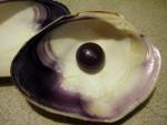
Fossil blister pearls attached to the shells of an Anodonta mollusk from China, early Early Pleistocene, are reported here for the first time. The pearls were investigated in detail using a variety of methods. Micro-CT scanning of the fossil pearls was carried out to discover the inner structure and the pearl nucleus. Using CTAn software, changes in the gray levels of the biggest pearl, which reflect the changing density of the material, were investigated. The results provide us with some clues on how these pearls were formed. Sand grains, shell debris or material with a similar density could have stimulated the development of these pearls. X-ray diffraction analysis of one fossil pearl and the shell to which it was attached reveals that only aragonite exists in both samples. The internal structures of our fossil shells and pearls were investigated using a Scanning Electron Microscope. These investigations throw some light on pearl development in the past.
The Nihewan beds, located in Hebei Province, northern China, are famous for their continuous deposition and complete set of Quaternary strata. Numerous studies were carried out after the “Nihewan Layer” was chosen as the Standard Stratum for the early Pleistocene of northern China (e.g. 1–4). Fossils excavated in the Nihewan area are both numerous and highly diverse, including human remains (e.g. 1, 4, 5), pollen and spores (e.g. 6–7), mammalian fossils (e.g. 8–10) and mollusks 11. In the course of collecting mollusk fossils in the Taiergou section, Nihewan area, the fossil pearls studied in this contribution were found by chance.
Very extensive and comprehensive article found here: http://journals.plos.org/plosone/article?id=10.1371/journal.pone.0164083
Join in and write your own page! It's easy to do. How? Simply click here to return to Pearl News.


